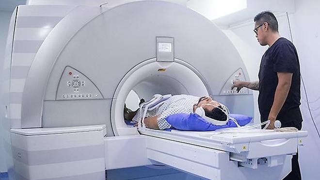
Spine imaging is often seen as the fastest way to find the cause of back pain, but a scan is not automatically the best first step. X-rays, CT scans and MRI can reveal fractures, infections, tumours, nerve compression and other important abnormalities. At the same time, they can also uncover changes that are common in healthy adults and may have little or nothing to do with a person’s pain.
This creates an important clinical dilemma. A patient may undergo an imaging test hoping for a clear explanation, only to be told that there is a disc bulge, degeneration or another abnormality. The finding can sound frightening even when it is not dangerous and does not require treatment.
The central question, therefore, is not simply whether a spine scan can detect something. Modern imaging can detect a great deal. The more useful question is whether the information from the scan is likely to change what happens next.
For uncomplicated back pain without warning signs, doctors may initially recommend appropriate activity, pain management, physiotherapy or observation rather than immediate imaging. Imaging becomes more important when symptoms, examination findings or medical history raise concern for a condition in which seeing the spine would alter treatment.
Why Doctors Do Not Automatically Scan Every Backache
Back pain is extremely common, and many episodes improve without invasive treatment. If every person with new back pain received an immediate scan, imaging would identify numerous structural changes that might never have caused symptoms.
Ageing naturally changes the spine. Discs can lose hydration and height. Joints can develop arthritic changes. Small disc bulges can appear even in people who do not experience pain.
These findings are real, but their presence does not automatically establish causation.
A scan shows anatomy; it does not always explain symptoms.
This distinction is one of the most important things patients should understand before undergoing spine imaging.
What Does an X-Ray Show?
A conventional X-ray is particularly useful for looking at bones and the overall alignment of the spine. It can help identify fractures, significant changes in bone structure, deformity and certain forms of arthritis.
However, an X-ray does not provide the same detailed view of nerves, spinal discs, the spinal cord or other soft tissues that MRI can provide.
Doctors may consider an X-ray when there is a reason to suspect a bony problem, particularly after trauma or when certain clinical circumstances make a fracture more plausible.
For ordinary nonspecific lower back pain, however, an X-ray may not provide information that changes management.
When Might a CT Scan Be Necessary?
CT, or computed tomography, provides much more detailed images of bone than a standard X-ray. It uses multiple X-ray measurements to create cross-sectional images and can be especially useful when doctors need to assess complex fractures or other detailed bony abnormalities.
CT can be valuable after significant trauma when a spinal fracture is suspected. It may also be used in situations where detailed bone anatomy is important for planning treatment.
However, CT uses ionising radiation. That does not mean CT should be avoided when medically necessary, but it does mean that the test should have a clear clinical purpose.
When the main question concerns spinal nerves, discs, the spinal cord or other soft tissues, MRI is often more informative.
When Is an MRI More Useful?
MRI uses magnetic fields and radio waves rather than ionising radiation. It provides detailed images of soft tissues and is particularly useful for evaluating the spinal cord, nerve roots, discs and surrounding structures.
Doctors may consider MRI when symptoms suggest significant nerve involvement or when there is concern about conditions such as infection, cancer or certain forms of spinal cord or nerve compression.
MRI can also become important when symptoms persist despite appropriate conservative management and the results could influence a decision about further treatment.
But even MRI has limitations.
A highly detailed scan can reveal abnormalities that are not responsible for the patient’s symptoms.
The Problem With “Abnormal” Spine Findings
One of the biggest misconceptions about medical imaging is that every abnormal finding is a disease.
Consider a person with back pain who undergoes an MRI and is told that they have degenerative disc disease. The phrase can sound alarming. Yet degenerative changes are common as people get older and may occur without significant symptoms.
Similarly, a disc bulge on an MRI does not automatically mean that the disc is the source of pain.
The challenge for doctors is therefore to interpret the scan alongside the patient’s symptoms and physical examination.
The image should support the clinical diagnosis, not replace it.
Incidental Findings Can Change How Patients Think About Their Bodies
Imaging can sometimes create a psychological problem even when the scan does not reveal a dangerous condition.
A patient who hears that their spine is “damaged” may become afraid to bend, exercise, walk or return to normal activities. That fear can alter behaviour and potentially contribute to prolonged disability.
In reality, many structural changes seen on spine imaging represent ordinary wear and ageing rather than evidence that the spine is fragile.
This is why communication matters as much as the scan itself.
A finding should be explained in context: Is it common? Does it match the symptoms? Is it causing nerve compression? Does it change treatment?
Why More Imaging Does Not Always Mean Better Care
It is tempting to assume that more information must produce a better medical decision.
But imaging can sometimes create a chain of events that begins with a relatively harmless finding.
A patient experiences back pain. A scan reveals a disc abnormality. The patient becomes worried. Additional consultations or procedures follow. Yet the original finding may not have been the true source of the symptoms.
This does not mean that surgery or other interventions are unnecessary in general. There are patients for whom imaging is essential to identifying a problem that genuinely requires specialist treatment.
The point is that the value of imaging depends on whether the result changes management in a meaningful way.
Red Flags: When Back Pain Needs More Urgent Assessment
The decision not to image immediately applies primarily to uncomplicated back pain. It should not be interpreted as a rule that imaging is unnecessary whenever someone has back pain.
Certain symptoms and medical circumstances can raise concern for a more serious underlying condition.
- Significant or high-impact trauma, particularly when a fracture is possible.
- New or progressive weakness in an arm or leg.
- Major changes in bladder or bowel control.
- Numbness involving the area around the groin or buttocks.
- Symptoms suggesting serious spinal cord or nerve compression.
- Fever or other evidence suggesting a significant infection, particularly with relevant risk factors.
- A history of cancer combined with concerning new symptoms.
- Unexplained systemic illness or other findings that make a serious underlying cause more likely.
- Severe or progressively worsening symptoms that do not fit a routine mechanical back-pain pattern.
These warning signs do not mean that a serious condition is definitely present. They mean that medical evaluation should not be delayed simply because uncomplicated back pain often improves without imaging.
X-Ray vs CT vs MRI: Which Scan Is Best?
| Imaging test | What it does best | Common reasons it may be considered | Key limitation |
|---|---|---|---|
| X-ray | Shows bones, alignment and certain structural changes | Suspected fracture, deformity or selected bony problems | Limited information about nerves, discs and soft tissues |
| CT scan | Provides detailed cross-sectional images, especially of bone | Complex fractures, trauma and detailed bony assessment | Uses ionising radiation |
| MRI | Shows discs, nerves, spinal cord and other soft tissues in detail | Suspected nerve compression, infection, cancer or selected persistent symptoms | Can reveal incidental findings that may not explain symptoms |
Why MRI Is Not Automatically the “Best” Scan
MRI is often perceived as the gold standard because it produces highly detailed images without ionising radiation.
But “most detailed” does not necessarily mean “most appropriate.”
If a doctor is trying to determine whether a recent injury has caused a complex fracture, CT may answer the question more effectively. If the clinical concern is a nerve or spinal cord problem, MRI may be more appropriate. If there is no indication of a serious structural problem, neither test may be necessary at the beginning.
The right scan is the one that answers the right clinical question.
Back Pain and Ageing: Why Scan Reports Can Be Misleading
Spinal degeneration is part of normal ageing for many people.
As the years pass, discs and spinal joints undergo structural changes. These may appear on imaging even when a person has no significant pain.
This creates a common interpretation trap.
If a patient has pain and the MRI shows degeneration, it is easy to assume that the degeneration caused the pain. But the scan alone cannot always establish that relationship.
The doctor must consider the location and character of the pain, neurological symptoms, examination findings and whether the imaging abnormality corresponds to the clinical picture.
In other words, correlation is more useful than simply finding something abnormal.
Does a Disc Bulge Mean You Need Surgery?
No.
A disc bulge or protrusion on imaging does not automatically mean that surgery is required.
Many disc abnormalities can be managed without an operation, depending on symptoms and clinical findings.
Surgery may become appropriate in selected patients when there is significant nerve compression, progressive neurological impairment or persistent symptoms that meet specific clinical criteria.
The important point is that the decision is based on the whole clinical picture rather than the presence of a disc abnormality alone.
Why Patients Often Ask for a Scan
For someone living with pain, uncertainty can be more frightening than the pain itself.
A patient may understandably think: “If we can see the spine, we will know exactly what is wrong.”
Sometimes that is true.
But imaging cannot always distinguish between a structural change that is incidental and one that is responsible for symptoms.
Patients should therefore feel comfortable asking their doctor a few practical questions before agreeing to a scan.
- What are we looking for?
- Will the result change my treatment?
- Why is this imaging test preferable to another one?
- What happens if the scan shows an abnormality that is unrelated to my pain?
- Are there warning signs that make imaging particularly important in my case?
These questions do not mean a patient is refusing care. They are part of informed decision-making.
The Hidden Cost of Unnecessary Imaging
The cost of unnecessary imaging is not limited to the price of the test.
There can be indirect financial costs, time spent attending appointments and the emotional burden of receiving a frightening-sounding report.
There can also be downstream consequences if incidental findings lead to unnecessary repeat imaging, procedures or specialist consultations.
At a healthcare-system level, avoiding low-value testing can allow resources to be directed towards patients who genuinely need advanced imaging and specialist treatment.
That is why appropriate imaging is not about doing less medicine. It is about doing the right medicine at the right time.
When Imaging Can Actually Change the Treatment Plan
The strongest reason to obtain a scan is that the result has the potential to alter what happens next.
For example, if a doctor suspects a serious infection or malignancy, imaging may help identify the problem and guide urgent management.
After significant trauma, imaging may reveal a fracture that requires immobilisation, specialist assessment or another intervention.
When neurological symptoms suggest substantial nerve or spinal cord involvement, MRI can provide information that may be critical to treatment decisions.
In these situations, delaying appropriate imaging can be more harmful than obtaining it.
A Better Way to Think About Spine Imaging
Instead of asking, “Do I need an MRI because my back hurts?” a more useful question is:
“Is there a specific clinical question that this scan can answer and that would change my treatment?”
This approach moves the conversation away from technology and towards clinical reasoning.
It also explains why two people with apparently similar back pain may receive completely different recommendations.
One person may need urgent imaging because of neurological deficits or trauma. Another may benefit more from conservative treatment and reassessment.
The Bottom Line
Spine imaging is an important medical tool, but it is not a universal answer to back pain. X-rays are useful for selected bone and alignment problems, CT provides detailed information about bone and complex injuries, and MRI is particularly valuable for nerves, discs, the spinal cord and other soft tissues.
The danger lies in assuming that every abnormality found on a scan is the cause of pain or evidence of serious spinal damage.
Many imaging findings can occur as part of normal ageing. When those findings do not match the patient’s symptoms and do not change treatment, they can create unnecessary anxiety rather than useful medical information.
The better strategy is targeted imaging: scan when the clinical situation provides a good reason, choose the modality that answers the specific question, and interpret the result alongside the patient’s symptoms and examination.
For people with uncomplicated back pain and no warning signs, the absence of an immediate scan does not mean their pain is being ignored. In many cases, it means the doctor is avoiding a test that is unlikely to improve the initial treatment plan.
And when genuine red flags appear, appropriate imaging becomes not an unnecessary expense, but a potentially crucial part of finding the right diagnosis and treatment.
For breaking news and live news updates, like us on Facebook or follow us on Twitter and Instagram. Read more on Latest Health on thefoxdaily.com.


COMMENTS 0