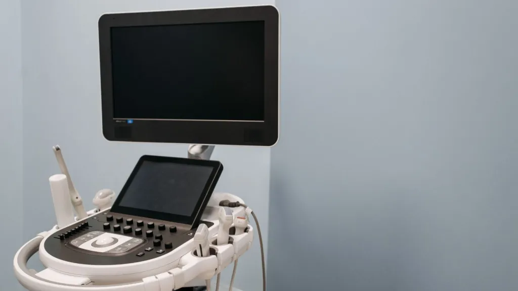
A handheld ultrasound device could help improve breast cancer detection in communities where access to specialist imaging and diagnostic facilities remains limited. An Indian study published in JCO Global Oncology has found that portable ultrasound, when operated by trained non-radiologist clinicians, can identify suspicious breast abnormalities with high sensitivity while reducing referrals for further assessment by approximately 50%.
The findings are significant for rural and underserved communities, where women may have to travel long distances to access mammography, radiologists and cancer specialists. By bringing preliminary imaging closer to patients, the approach could help healthcare workers identify women who need further investigation while reducing unnecessary referrals among those whose abnormalities are likely to be benign.
The study evaluated portable ultrasound as a triage tool following a clinical breast examination (CBE). It reported an overall sensitivity of 94.3% and specificity of 76.7% for detecting breast cancer. Researchers also found that adding ultrasound improved the positive predictive value of clinical breast examinations.
Although the technology is not intended to replace mammography, biopsy or specialist diagnosis, the findings suggest that portable imaging could become a useful part of early detection programmes, particularly in healthcare systems with limited diagnostic resources.
How a handheld ultrasound helped a woman in Kerala detect breast cancer
The potential impact of portable ultrasound is illustrated by the experience of Noorjehan, whose name has been changed to protect her identity. Three years ago, the 53-year-old attended a breast cancer screening camp in Chakkarakkal, near Kannur in Kerala. Like several women in her village, she had been hesitant to seek medical attention because of limited resources and fear of what a diagnosis might reveal.
Her clinical breast examination produced a positive finding, prompting healthcare workers to conduct a portable ultrasound assessment. The imaging was performed by clinicians trained by the International Agency for Research on Cancer (IARC) at the Malabar Cancer Care Society (MCCS).
The ultrasound assessment helped determine that she needed additional investigation. Noorjehan was referred for mammography and other confirmatory tests, which established that she had breast cancer. She was subsequently referred to a local medical college, where she underwent surgery followed by chemotherapy.
By September 2024, she had completed her treatment and recovered. The MCCS team has continued to follow up with her.
Dr D K Pai, president and founder of MCCS and a senior author of the study, said the screening initiative helped bring diagnostic services within reach of women who might otherwise struggle to access them.
He explained that diagnostic facilities were neither easily accessible nor affordable for Noorjehan’s family. Without detection, her cancer could have progressed to a more advanced stage, making treatment more complicated and expensive.
“The screening camp at her doorstep saved her,” Dr Pai said.
Her experience highlights why access to early detection matters. Identifying breast cancer before it advances can give patients an opportunity to receive timely treatment. For women living far from hospitals or specialist centres, community-based screening can also reduce some of the practical barriers that delay diagnosis.
What is portable ultrasound and how does it work?
Portable ultrasound is a compact medical imaging system that uses high-frequency sound waves to create images of internal body tissues. Unlike conventional ultrasound equipment commonly found in hospitals and diagnostic centres, handheld devices are lightweight and can often operate on batteries.
Many models connect to a smartphone or tablet, allowing healthcare workers to view images without relying on large imaging machines. Their portability makes them particularly useful in community health programmes, mobile clinics and locations where access to conventional diagnostic infrastructure is limited.
In breast screening, portable ultrasound provides information that a physical examination alone cannot offer. During a clinical breast examination, a healthcare worker uses touch to identify lumps or other abnormalities. However, this method cannot produce an image of the underlying breast tissue.
Portable ultrasound adds an imaging component to the assessment. Trained clinicians can examine an abnormality and look for features that help determine whether it is likely to require further investigation.
The purpose in this programme is not to conduct a complete diagnostic examination independently. Instead, the device is used to answer a limited set of clinical questions and help guide decisions about which women should be referred for additional testing.
This distinction is important because portable ultrasound is being evaluated as a triage tool rather than a replacement for specialist breast imaging. Women with suspicious findings still require appropriate diagnostic investigations to establish whether cancer is present.
How IARC is training non-radiologist clinicians to use handheld ultrasound
A major feature of the initiative is that portable ultrasound can be operated by trained clinicians who are not specialist radiologists. This could help address one of the central challenges facing breast cancer detection programmes in low-resource settings: the shortage or limited availability of specialist imaging professionals.
The International Agency for Research on Cancer has been training non-radiologist clinicians and healthcare workers to conduct preliminary breast ultrasound assessments. The training combines online learning modules with short, practical clinical workshops.
Participants learn how to operate the portable equipment, obtain images and interpret selected features relevant to the screening process. The training is designed around a focused clinical task rather than the broader range of examinations performed by specialist radiologists.
This approach could allow trained frontline healthcare workers to provide an additional level of assessment at community screening camps and primary healthcare facilities. Instead of referring every woman with an abnormal clinical examination to a distant diagnostic centre, healthcare teams can use preliminary imaging to help prioritise referrals.
However, the quality of the assessment depends on appropriate training, practical experience, supervision and clear referral protocols. Portable devices alone cannot guarantee accurate results. The people using them must understand their limitations and know when a patient requires further evaluation.
Study finds 94.3% sensitivity and 50% fewer referrals
The Indian study evaluated portable ultrasound triage in 319 women aged 40 to 75 years. Participants came from two healthcare settings: community-based screening programmes involving women without symptoms and diagnostic clinics catering to women who had breast-related symptoms.
Trained non-radiologist clinicians used portable ultrasound to assess abnormalities identified during clinical breast examinations. The objective was to distinguish findings that appeared suspicious and required further investigation from those more likely to be benign.
The results showed an overall sensitivity of 94.3% and specificity of 76.7% for breast cancer detection.
These two measures describe different aspects of a screening or diagnostic assessment:
- Sensitivity: The proportion of people who have the disease and are correctly identified by the test. In this study, the reported sensitivity was 94.3%.
- Specificity: The proportion of people who do not have the disease and are correctly identified as negative. The study reported a specificity of 76.7%.
- Positive predictive value: The proportion of people with a positive test result who actually have the disease. The researchers found that portable ultrasound improved this measure when added to clinical breast examination.
One of the most important findings was the reduction in referrals. The use of portable ultrasound following clinical breast examination reduced referrals for further assessment by approximately 50%.
This matters because not every lump or abnormality detected during a physical examination is cancerous. Without access to preliminary imaging, women with benign findings may still be referred to larger hospitals for additional tests. Such referrals can involve travel expenses, lost working hours, anxiety and additional costs for families.
By helping clinicians identify which abnormalities are more likely to need further investigation, portable ultrasound could make screening pathways more efficient. It may also allow specialist diagnostic facilities to concentrate their resources on women who require more detailed assessment.
Why reducing unnecessary referrals matters for rural healthcare
Access to breast cancer diagnosis is not determined by the availability of screening camps alone. Women also need a clear and affordable pathway from an initial examination to confirmatory tests and treatment.
In many resource-constrained settings, a woman with a suspicious clinical breast examination may need to travel to a district hospital, medical college or specialised cancer centre for imaging. These journeys can be difficult for people living in remote villages, particularly when transport costs, family responsibilities and lost wages are involved.
There is also the psychological burden of waiting for further tests. A breast lump can cause considerable anxiety, even though many breast abnormalities are not cancerous. When diagnostic services are far away, uncertainty can continue for longer and may discourage some women from completing the referral process.
Portable ultrasound could help address part of this problem by providing an additional assessment closer to the community. If the findings suggest that an abnormality is unlikely to be malignant, the healthcare team may be able to avoid an unnecessary referral. If the findings are suspicious, the woman can be directed towards confirmatory investigations.
The reduction in referrals reported by the study therefore has implications beyond convenience. It could help improve the use of limited specialist resources while making the screening process less burdensome for patients.
However, fewer referrals should not become the primary measure of success. Screening programmes must ensure that women with possible cancer are not incorrectly reassured or lost during follow-up. Reliable referral systems and access to confirmatory testing remain essential.
Can portable ultrasound help improve breast cancer screening in India?
India faces the challenge of delivering timely cancer detection across communities with very different levels of healthcare access. Large hospitals may offer specialist imaging and treatment, but women in smaller towns and rural areas can encounter difficulties reaching these services.
Community-based screening programmes can help identify women who need further assessment, but their effectiveness depends on what happens after an abnormality is detected. A screening examination without an accessible diagnostic pathway may not deliver the intended benefit.
Portable ultrasound offers a possible way to strengthen this pathway. Because the equipment can be transported and used outside conventional imaging departments, trained healthcare workers may be able to conduct preliminary assessments during community outreach programmes.
The approach could be particularly relevant in areas where mammography facilities and specialist radiologists are not readily available. It may also help healthcare organisations organise referrals more effectively by identifying patients who need additional investigations.
Dr Partha Basu, Head of the Early Detection, Prevention, and Infections Branch at IARC and a co-author of the study, said the findings indicate that portable ultrasound could improve the efficiency of early detection programmes where access to specialist imaging is limited.
The study provides encouraging evidence, but wider implementation would require careful planning. Healthcare systems would need to invest in training, equipment maintenance, image quality checks, supervision and referral arrangements. They would also need to ensure that women who receive suspicious results can access mammography, biopsy and treatment without unnecessary delays.
Artificial intelligence could strengthen portable breast ultrasound
The research team is also exploring how artificial intelligence could support the use of portable ultrasound in low-resource settings. According to Dr Basu, images collected through the project have already been used to train and test an AI algorithm designed to detect breast cancers.
AI-assisted analysis could potentially help clinicians interpret ultrasound images by identifying patterns associated with suspicious abnormalities. This may be particularly useful in healthcare settings where experienced imaging specialists are not always available.
Combining portable ultrasound with AI could eventually provide frontline healthcare workers with additional decision-support tools. Instead of relying entirely on manual interpretation, clinicians may be able to use software-generated assessments alongside their training and clinical judgement.
However, AI should be viewed as a potential support system rather than an independent replacement for medical professionals. Algorithms need to be tested across diverse patient populations and different healthcare environments. Their performance also depends on the quality and representativeness of the images used during development.
Before such tools can become part of routine screening, researchers and healthcare authorities will need to establish how accurately they perform in real-world conditions and whether they improve patient outcomes.
Can handheld ultrasound replace mammography?
Despite the encouraging results, portable ultrasound should not be considered a replacement for mammography or other established diagnostic procedures. The study evaluated it as a triage tool used after clinical breast examination, with the aim of identifying women who require further assessment.
Mammography remains an important breast imaging method, while ultrasound can provide additional information about breast abnormalities. When a finding is suspicious, further investigations may include specialist imaging and a biopsy to establish whether cancer is present.
Ultrasound also has limitations. Its accuracy can depend on the operator’s training, the quality of the equipment and the characteristics of the abnormality being examined. A negative preliminary assessment does not eliminate the need for further evaluation when symptoms or clinical findings remain concerning.
For this reason, portable ultrasound is best understood as one component of a wider early detection system. Its value lies in bringing preliminary imaging closer to women and helping healthcare workers make better-informed referral decisions.
What the findings mean for the future of early breast cancer detection
The Indian study highlights how relatively compact medical technologies can help address gaps in healthcare delivery when combined with appropriate training and referral systems. Portable ultrasound does not remove the need for specialist diagnosis, but it may help make the first stages of breast cancer assessment more accessible.
The experience of Noorjehan demonstrates the importance of taking screening services closer to communities where cost, distance and fear can prevent women from seeking medical attention. Her diagnosis led to further testing and treatment, showing how a community-based approach can connect patients with the care they need.
The study’s reported sensitivity of 94.3% and approximately 50% reduction in referrals provide encouraging evidence for further evaluation of the model. Its potential impact could be greater when portable imaging is combined with trained frontline clinicians, reliable follow-up systems and carefully validated AI-assisted tools.
For wider adoption, however, healthcare programmes must ensure that screening is linked to affordable confirmatory diagnosis and timely treatment. Detecting a suspicious abnormality is only the beginning of the process; patients must be able to complete the next steps in their care.
Ultimately, the promise of handheld ultrasound lies in making breast cancer assessment more accessible without compromising the quality of diagnosis. For women in underserved communities, bringing a useful imaging tool closer to home could help reduce barriers to care and strengthen the chances of identifying cancer early.
For breaking news and live news updates, like us on Facebook or follow us on Twitter and Instagram. Read more on Latest Health on thefoxdaily.com.






COMMENTS 0