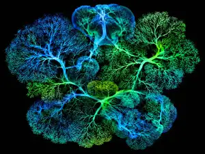
Researchers associated with the Massachusetts Institute of Technology (MIT) have created one of the most detailed maps yet of how a mammal develops, tracing the family history of more than 1.4 million cells in developing mouse embryos. The study offers an unusually comprehensive view of how cells divide, change their genetic activity and gradually acquire the identities needed to build different parts of the body.
Every cell in an animal ultimately originates from a fertilised egg. As that original cell divides, its descendants multiply and develop into specialised cell types that form organs, tissues and other structures. Although scientists have studied individual stages of embryonic development for decades, following the relationships between such a large number of cells across the developing embryo has remained challenging.
The new research brings together cellular family trees, gene activity and information about where cells are located. This allows scientists to investigate not only what a cell eventually becomes but also how early its developmental path begins to narrow towards a particular fate.
The study was led by researchers in the laboratory of MIT professor Jonathan Weissman, who is also associated with the Whitehead Institute for Biomedical Research. The work involved collaboration with researchers at Yale University and the Max Planck Institute for Molecular Genetics and was published in the journal Cell on October 8, 2026.
How scientists traced the family history of millions of embryonic cells
To understand how cells develop into different parts of an organism, researchers need more than a picture of an embryo at a particular moment. They need to know which cells descended from which earlier cells and how those relationships changed as development progressed.
The MIT team’s approach involved reconstructing cellular lineage trees. Similar to a family tree that records relationships between generations of people, a cell lineage tree shows how individual cells are related through successive rounds of division.
Researchers used a technology called PEtracer, which introduces small, heritable genetic marks into cells as development progresses. These marks are passed on when a cell divides, creating a record that can later be examined to reconstruct the relationships between descendants.
By analysing these genetic records, scientists can work backwards to identify which cells share common ancestors. This provides information about the developmental history of cells that would be difficult to obtain by observing an embryo at only one point in time.
The team combined the reconstructed family trees with gene-expression information and the locations of cells within the embryos. Gene expression refers to the activity of genes that cells use to carry out their functions. Different patterns of gene activity help determine whether a cell develops into a particular specialised type.
This combination is important because lineage alone cannot explain every aspect of cell development. Two cells may share an ancestor but eventually develop into different types of tissue. By examining ancestry alongside gene activity and location, researchers can investigate when cells begin following different developmental paths.
Study analysed more than 1.4 million cells from 16 mouse embryos
The scale of the research is one of its most significant features. Scientists analysed more than 1.4 million cells from 16 mouse embryos collected at multiple stages of early development.
The embryos covered the period from embryonic day 7.5 to embryonic day 10.0, with samples collected at half-day intervals. This period includes important stages of gastrulation and early organ formation, when cells begin organising into the structures that will eventually develop into the animal’s body.
The researchers reconstructed approximately 75 per cent of cell divisions across the analysed dataset. This allowed them to build a detailed picture of how cellular lineages develop and how different groups of cells contribute to particular tissues.
Importantly, the study did not simply produce a collection of images showing different developmental stages. It created a resource that connects cellular ancestry with molecular information, helping researchers examine the relationships between early cell divisions and later cell identities.
The team also found that lineage patterns were strikingly reproducible across independent embryos. This suggests that, despite the flexibility involved in mammalian development, many aspects of the way cells become organised follow consistent patterns.
When do embryonic cells begin developing into different tissues?
One of the central questions in developmental biology is when cells begin committing to particular identities. In the earliest stages of development, cells have the potential to contribute to several different parts of the embryo. As development progresses, their possible destinations become increasingly restricted.
This process is known as cell fate specification. It describes how a developing cell acquires the characteristics that guide it towards becoming a particular type of cell.
For example, the developing embryo must generate cells that eventually contribute to the nervous system, muscles, blood vessels, skin and internal organs. These specialised cells do not appear independently. They emerge through a series of divisions and developmental changes involving earlier cell populations.
The new lineage map allows scientists to examine how early ancestors contribute to different groups of specialised cells. It also helps identify when certain developmental possibilities become more restricted.
Rather than treating the formation of each organ as an isolated process, researchers can study how different developmental pathways are connected. This provides a wider view of how the embryo develops as an integrated system.
Research reveals new details about neural crest and blood vessel development
The study identified several specific patterns in the development of different tissues. One finding involved the progressive restriction of neural crest cell fate.
Neural crest cells are a specialised group of embryonic cells that contribute to several structures in the developing body. Their descendants help form parts of the peripheral nervous system and other tissues. Understanding how these cells acquire increasingly specialised identities is important for explaining how complex structures emerge during development.
The researchers’ lineage analysis provided a way to examine how neural crest developmental potential becomes progressively restricted. This helps scientists understand the relationship between early cell ancestry and the specialised roles cells eventually perform.
Another area examined by the team involved the formation of endothelial cells, which make up the inner lining of blood vessels.
The study investigated the relative contributions of different mesodermal origins to the development of these cells. Mesoderm is one of the primary embryonic tissue layers and contributes to structures such as muscles, blood vessels and connective tissues.
Understanding which early cell populations contribute to endothelial cells can help researchers investigate how the vascular system develops and how different developmental pathways contribute to its formation.
The researchers also examined the dynamics of axial elongation, a process through which the developing embryo extends along its main body axis. This process is essential for establishing the overall shape and organisation of the body.
How the cell lineage map could help scientists understand birth defects
One of the important potential applications of the research is improving the understanding of birth defects. Many developmental abnormalities occur when cells do not follow the expected pathways while tissues and organs are being formed.
Normal embryonic development requires several processes to happen in a coordinated manner. Cells must divide, move, communicate and activate the appropriate genes at particular stages. If a critical process is disrupted, the resulting tissue or organ may not develop as expected.
A detailed map of normal development could help researchers identify where these processes begin to go wrong. By comparing developmental patterns, scientists may be able to investigate whether an abnormality is associated with an early change in cell fate, an altered lineage relationship or a difference in gene activity.
For example, if a group of cells that normally contributes to a particular tissue follows a different developmental path, researchers could use lineage information to investigate when that change occurred.
The study does not provide a direct diagnostic test or an immediate treatment for birth defects. Instead, it creates a research resource that could help scientists investigate the biological events behind developmental abnormalities.
Why the research could also matter for cancer studies
Although the study focuses on healthy mouse embryos, its findings could also contribute to cancer research.
Cancer develops when cells acquire changes that allow them to grow and behave differently from normal cells. Understanding the history of a cell can help scientists investigate how those changes emerge and how particular cell populations develop characteristics associated with disease.
The lineage-tracing approach used in the study offers a way to examine relationships between cells and reconstruct aspects of their developmental history. Researchers hope that similar principles can help investigate how cancer cells change in ways that allow them to spread or resist treatment.
Cell ancestry is relevant because the behaviour of a cell can be influenced by its developmental background. Two cells that appear similar at a particular moment may have different histories, while cells with a shared ancestor may eventually acquire very different characteristics.
A better understanding of these relationships could help researchers investigate how cancer cells evolve and why some cell populations behave differently from others.
However, the mouse embryo study does not demonstrate a new cancer treatment or establish that the same developmental patterns directly explain human tumours. Its significance lies in providing a more detailed framework for studying cell histories and biological changes.
Could the map improve stem cell research?
The research may also have implications for stem cell biology. Stem cells are important because they can develop into different specialised cell types under suitable conditions. Scientists are investigating ways to guide these cells towards becoming functional tissues for research and potential medical applications.
One major challenge is reproducing the conditions that naturally exist inside an embryo. In a developing organism, cells receive a combination of molecular signals and environmental information that influence what they become. Recreating the right sequence of signals in a laboratory can be difficult.
A detailed map of embryonic development could help researchers understand the sequence of changes that takes a cell from an early state to a specialised identity.
Such information could eventually help scientists design more precise methods for producing particular cell types from stem cells. Instead of relying only on trial and error, researchers could use developmental maps to investigate which molecular conditions are associated with specific cell-fate decisions.
This could be valuable for studies involving nerve cells, muscle cells, blood vessel cells and other specialised tissues. However, translating the findings into reliable medical applications will require further research.
How this research differs from traditional embryo studies
Traditional developmental biology has often relied on studying embryos at selected stages. Researchers might examine the developing heart, nervous system or another structure at a particular point and compare it with samples collected later.
These studies have provided essential information about how organs form. However, individual snapshots do not always reveal the complete history of the cells involved.
The new approach adds a lineage dimension. It allows researchers to connect cells observed at different developmental stages through their inherited genetic marks.
Combining this information with gene-expression data and cell locations creates a more detailed picture of the process. Scientists can investigate not only where a cell is found but also which ancestors contributed to it and how its molecular identity changed.
The resulting Mouse Embryonic Lineage Atlas provides researchers with a reference that can be explored to study cell types, lineage relationships and gene activity across early mouse development.
What the findings mean for future developmental biology research
The MIT-associated research represents a major step towards understanding mammalian development at the level of individual cells and their ancestry. By reconstructing more than 1.4 million cellular histories, the team has created a resource that connects early cell divisions with the formation of specialised tissues.
Its value extends beyond the number of cells analysed. The combination of lineage trees, gene activity and spatial information offers researchers a way to investigate developmental decisions that were previously difficult to study at such a large scale.
Future research could build on this foundation to examine how normal developmental pathways differ when genetic or environmental conditions change. It could also help scientists compare findings across different tissues and investigate how early cellular decisions influence later biological outcomes.
The findings are based on mouse embryos, so they should not automatically be treated as a complete map of human development. Mammalian development shares important biological principles, but differences between species mean that further studies will be needed to understand how directly the results apply to humans.
For now, the research provides a detailed reference for studying how a single fertilised egg gives rise to a complex organism. By turning millions of individual cellular histories into a connected developmental map, scientists are getting closer to understanding when cells begin choosing their identities and how those early decisions shape the formation of the body.
For breaking news and live news updates, like us on Facebook or follow us on Twitter and Instagram. Read more on Latest World on thefoxdaily.com.






COMMENTS 0